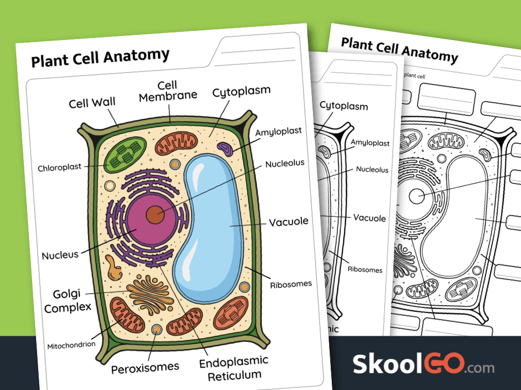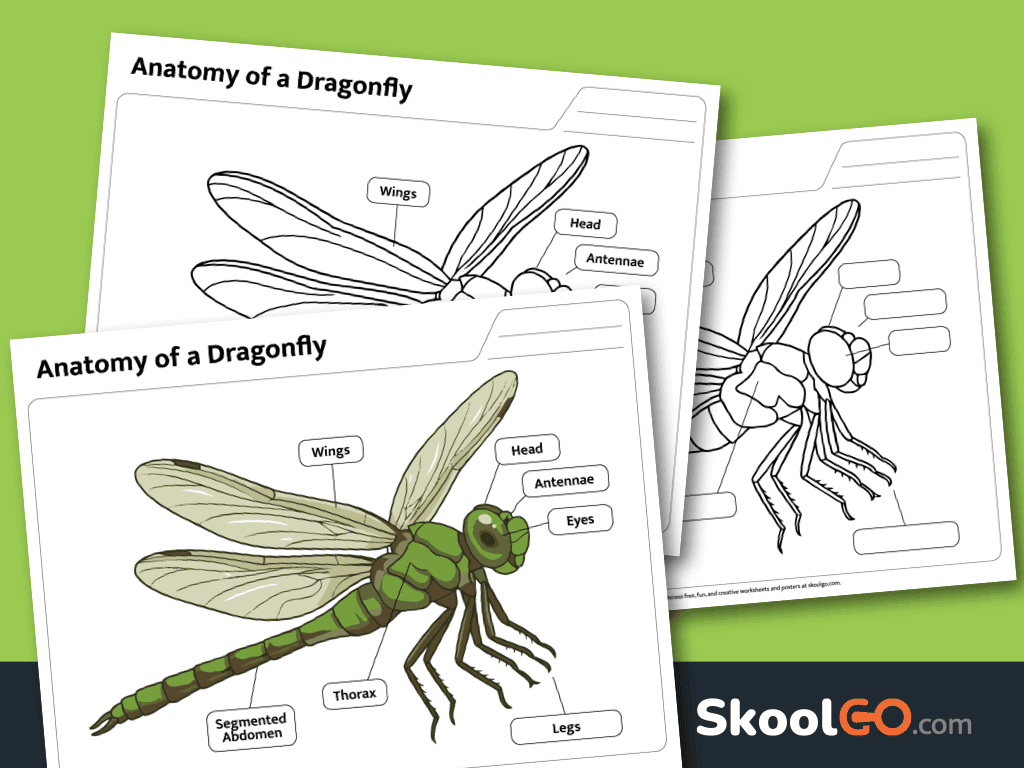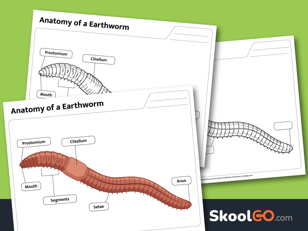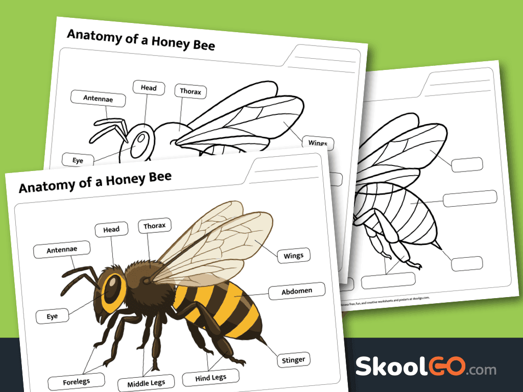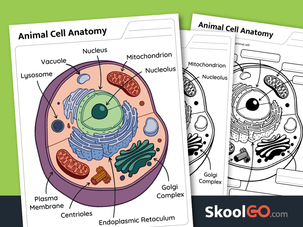
Animal Cell Anatomy Labeling Worksheet is a printable animal cell anatomy worksheet that helps learners identify key cell structures. Through observation and labeling, students build science vocabulary while developing a deeper understanding of animal cells.
Explore Animal Cell Structures Through Labeling
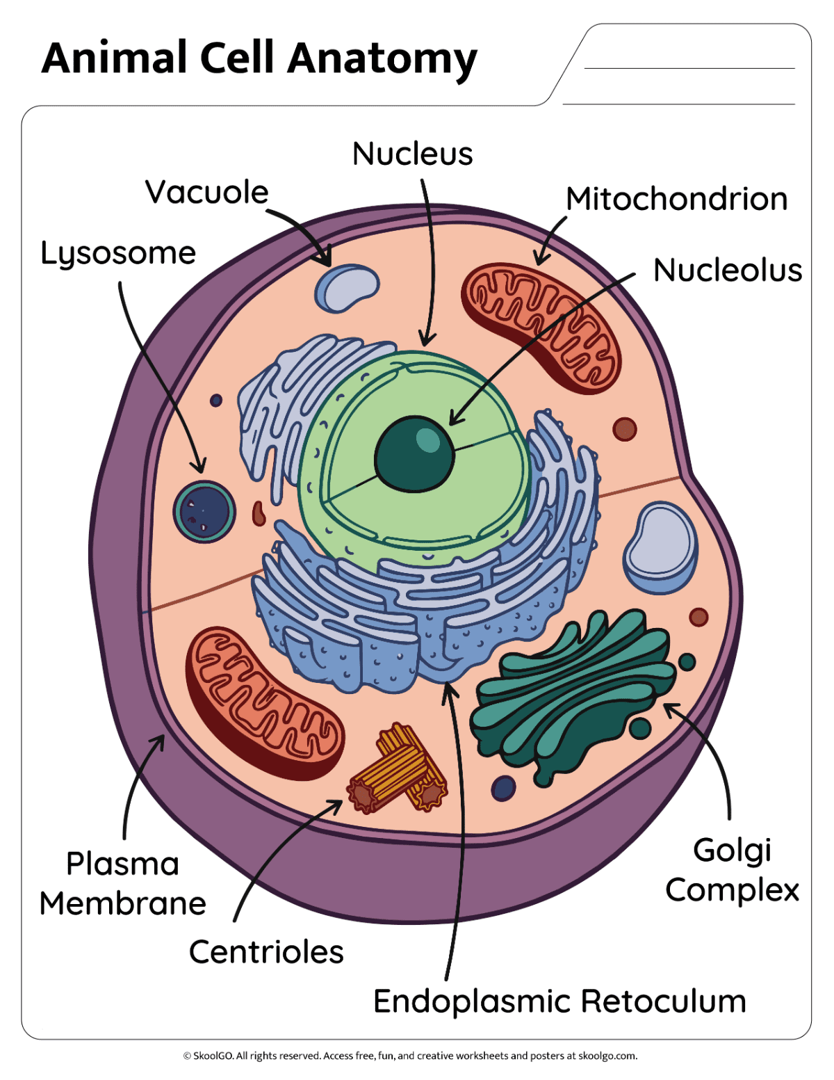
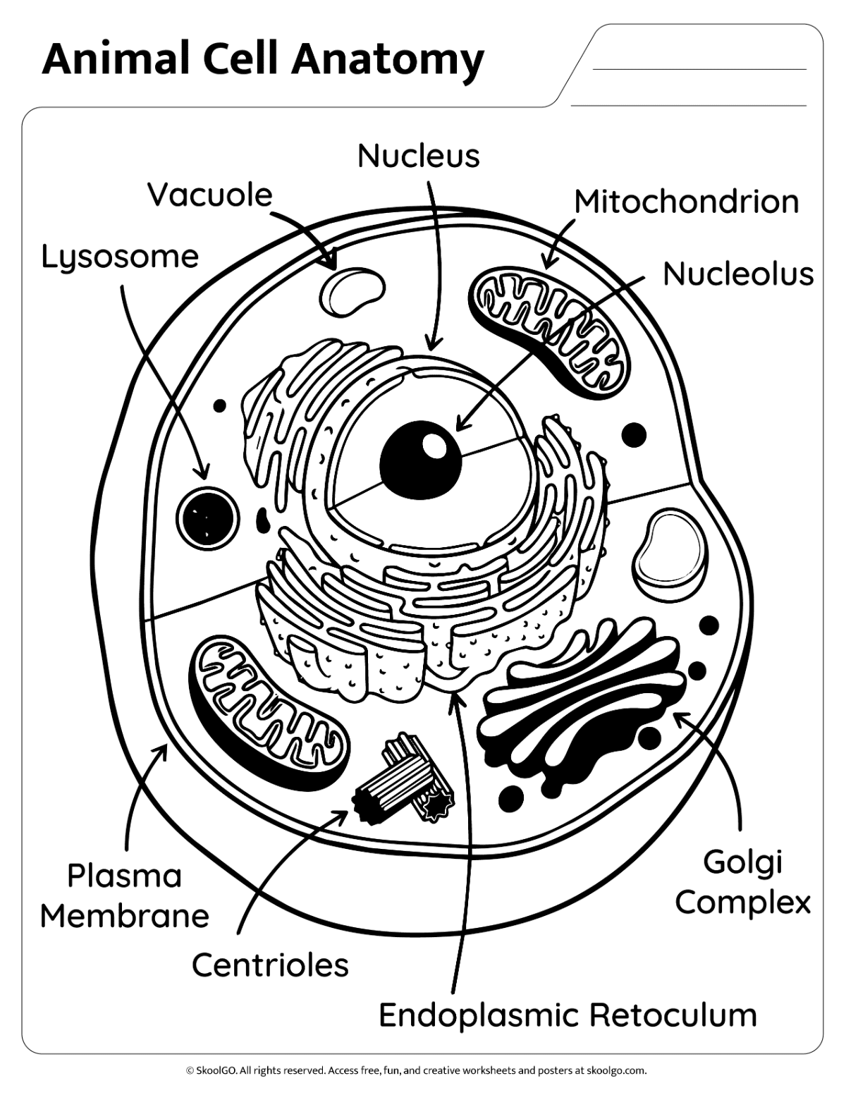
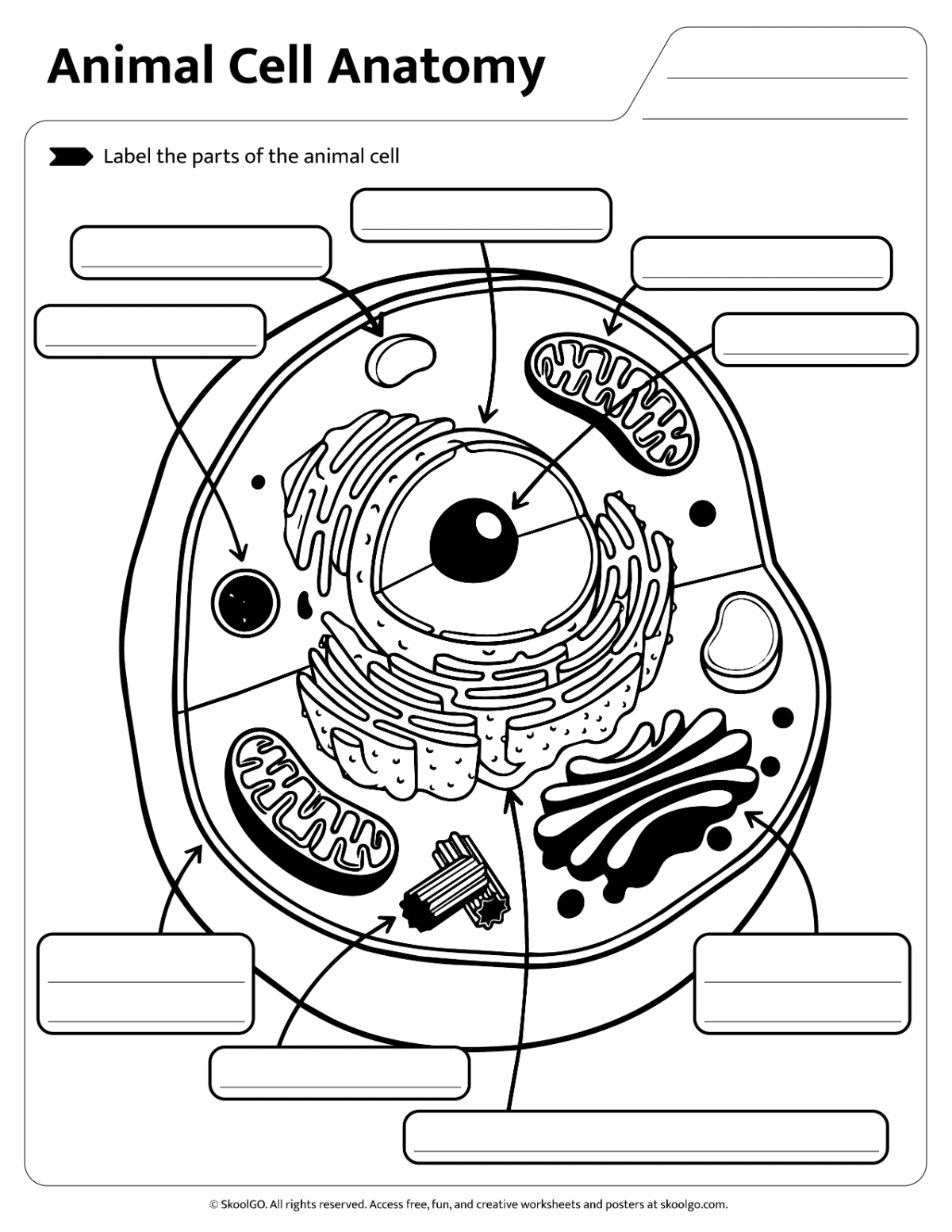
This resource features a detailed animal cell diagram with clearly illustrated organelles. Additionally, learners can study labeled examples before completing an independent labeling activity. The layout supports focused learning and encourages careful examination of each cell part.
Students strengthen vocabulary and observation skills while learning important biology concepts. Furthermore, the worksheet promotes visual perception and reading comprehension. As a result, learners become more confident when identifying structures such as the nucleus, mitochondrion, vacuole, and Golgi complex.
The document includes three pages. Firstly, learners review a full-color labeled diagram. Secondly, they can use a black-and-white labeled version for printing or coloring. Finally, they complete a labeling practice page. The resource uses portrait orientation, standard Letter size, and PDF format.
Download this worksheet to support science lessons, homeschool activities, or independent practice. Overall, it provides an engaging way to reinforce animal cell anatomy while encouraging active learning and scientific discovery.
Worksheet preview:
The worksheet document features:
-
- One full-color labeled animal cell anatomy diagram
- One black-and-white labeled animal cell anatomy diagram
- One animal cell labeling practice worksheet
- Unique and exclusive design
- Portrait orientation
- Standard Letter size (8.5 x 11 inches)
- Available as a downloadable PDF file